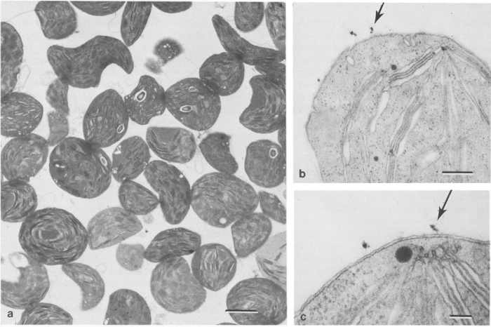
Label the transmission electron microscope image of a chloroplast below – Labeling the transmission electron microscope image of a chloroplast below unveils the intricate inner workings of this vital organelle. Through the lens of TEM, we embark on a journey to identify and comprehend the structures that orchestrate photosynthesis, the life-sustaining process that fuels our planet.
Chloroplasts, the powerhouses of plant cells, are the primary sites of photosynthesis. Understanding their structure and function is crucial for unraveling the mysteries of plant biology and advancing our knowledge of life on Earth.
Chloroplast Structure

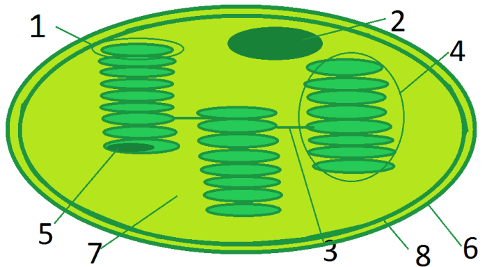
Chloroplasts are organelles found in plant cells that are responsible for photosynthesis. They have a double membrane structure, with the inner membrane forming flattened sacs called thylakoids. These thylakoids are stacked together to form grana, which are connected by stroma lamellae.
Arrangement and Functions of the Thylakoid Membranes
The thylakoid membranes contain chlorophyll and other pigments that absorb light energy. This light energy is used to split water molecules into hydrogen and oxygen. The hydrogen is then used to reduce carbon dioxide into glucose, while the oxygen is released as a waste product.
Detailed Illustration of the Chloroplast with Labeled Components
- Outer membrane:The outermost membrane of the chloroplast, which surrounds the entire organelle.
- Inner membrane:The membrane that lines the inside of the chloroplast and forms the thylakoids.
- Thylakoids:Flattened sacs that contain chlorophyll and other pigments.
- Grana:Stacks of thylakoids.
- Stroma lamellae:Membranes that connect the grana.
- Stroma:The fluid-filled space inside the chloroplast that contains enzymes and other molecules.
Transmission Electron Microscopy (TEM): Label The Transmission Electron Microscope Image Of A Chloroplast Below
Transmission electron microscopy (TEM) is a technique that uses a beam of electrons to create images of thin specimens. It is used to study the ultrastructure of cells and tissues.
Principle and Applications of TEM
TEM works by passing a beam of electrons through a thin specimen. The electrons interact with the specimen, and the resulting scattering and absorption of electrons creates an image. TEM is used to study the ultrastructure of cells and tissues, including the structure of chloroplasts.
Sample Preparation Process for TEM Imaging of Chloroplasts
To prepare chloroplasts for TEM imaging, they must be fixed, dehydrated, and embedded in a resin. The resin is then cut into thin sections, which are placed on a grid and examined under the TEM.
Overview of the Different Types of TEM Images and Their Uses
TEM can be used to create different types of images, including bright-field images, dark-field images, and high-resolution images. Bright-field images show the overall structure of the specimen, while dark-field images show the distribution of heavy atoms. High-resolution images can be used to resolve the fine details of the specimen.
Labeling the Chloroplast TEM Image

The TEM image of a chloroplast shows the overall structure of the organelle, including the outer membrane, inner membrane, thylakoids, grana, and stroma. The following table identifies the key features visible in the image:
| Structure | Function | Characteristics |
|---|---|---|
| Outer membrane | Surrounds the chloroplast | Smooth surface |
| Inner membrane | Forms the thylakoids | Folded into flattened sacs |
| Thylakoids | Contain chlorophyll and other pigments | Stacked together to form grana |
| Grana | Stacks of thylakoids | Connected by stroma lamellae |
| Stroma lamellae | Membranes that connect the grana | Contain enzymes and other molecules |
| Stroma | Fluid-filled space inside the chloroplast | Contains enzymes and other molecules |
Proper labeling of TEM images is important for accurate interpretation of the images. The labels should be clear and concise, and they should accurately describe the structures that are being shown.
Organelle Interactions

Chloroplasts interact with other organelles in the cell, including mitochondria and the endoplasmic reticulum. These interactions are important for cellular metabolism.
Interactions Between Chloroplasts and Mitochondria
Chloroplasts and mitochondria are the two main organelles involved in cellular respiration. Chloroplasts produce ATP through photosynthesis, while mitochondria produce ATP through oxidative phosphorylation. The ATP produced by chloroplasts is used to power the Calvin cycle, which is the light-independent reactions of photosynthesis.
The ATP produced by mitochondria is used to power the Krebs cycle, which is the citric acid cycle.
Interactions Between Chloroplasts and the Endoplasmic Reticulum
Chloroplasts and the endoplasmic reticulum (ER) are both involved in the synthesis of lipids. The ER synthesizes phospholipids, which are the main components of cell membranes. Chloroplasts synthesize galactolipids, which are a type of lipid that is found in the thylakoid membranes.
How TEM Can Be Used to Study Organelle Interactions, Label the transmission electron microscope image of a chloroplast below
TEM can be used to study the interactions between chloroplasts and other organelles. For example, TEM can be used to visualize the close association between chloroplasts and mitochondria, which is known as the chloroplast-mitochondria complex. TEM can also be used to visualize the transfer of lipids between chloroplasts and the ER.
Answers to Common Questions
What is the function of the thylakoid membranes in a chloroplast?
The thylakoid membranes house the chlorophyll molecules responsible for capturing light energy during photosynthesis.
How does TEM contribute to our understanding of chloroplast structure?
TEM provides high-resolution images that reveal the ultrastructure of chloroplasts, allowing us to visualize and characterize their intricate components.
Why is accurate labeling of TEM images important?
Proper labeling ensures precise identification and interpretation of the structures observed in TEM images, facilitating accurate scientific communication and understanding.